Our laboratory focuses on the development of plant-derived hydrocolloids as sustainable biomaterials for tissue engineering applications. We explore green, water-based extraction methods to obtain functional biopolymers with favorable gelation, swelling, and biocompatibility properties. These naturally sourced materials such as quince seed hydrocolloid, psyllium seed hydrocolloid, and flaxseed hydrocolloid, are processed into 3D scaffolds using environmentally friendly fabrication techniques. We systematically investigate their chemical, morphological, mechanical, and biological characteristics to support long-term cell culture and promote tissue regeneration.
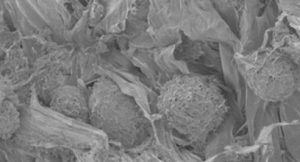
In our lab, we are harnessing the power of plants to create sustainable and biocompatible scaffolds for tissue engineering through plant decellularization. By carefully removing cellular components from plant tissues while preserving their intricate vascular architecture and cellulose-rich extracellular matrix, we obtain naturally derived scaffolds that are lightweight, porous, and structurally similar to human tissues. These decellularized plant matrices offer an eco-friendly and cost-effective platform for supporting cell adhesion, growth, and tissue regeneration. With their unique microstructure and versatility, plant-based scaffolds hold exciting promise for applications ranging from soft tissue repair to complex organ regeneration.
Animal-based polymers play a significant role in tissue engineering, particularly by decellularized tissues and extracellular matrix (ECM) components. These materials retain the native biochemical cues and structural architecture of the original tissue, providing an ideal scaffold for cell attachment, proliferation, and differentiation. Decellularization processes remove cellular components while preserving ECM proteins such as collagen, elastin, and glycosaminoglycans, making them biocompatible and immunogenically safer for implantation. The resulting ECM-based scaffolds are widely used in regenerative medicine applications, including neuronal, skin, cardiac, and musculoskeletal tissue repair, due to their ability to mimic the natural microenvironment and promote tissue regeneration.
We are actively engaged in the development of plant derived hydrocolloid bio-inks for 3D bioprinting applications. Our research focuses on hydrocolloids with tunable rheological and gelling properties, enabling low-pressure printability and stability in cell culture environments. By utilizing green extraction methods, we obtain plant-based bioinks such as Quince Seed Hydrocolloid and Psyllium Seed Hydrocolloid, which are suitable for scaffold fabrication by 3D bioprinting. These bioinks are systematically evaluated for their printability, structural integrity, and biocompatibility to support long-term cell viability and functionality.
In our lab, we are developing a novel biomimetic hybrid bioink composed of quince seed hydrocolloid (QSH), gelatin (Gel), and the self-assembling P11-4 peptide to support dental tissue engineering. The incorporation of P11-4 enhances the scaffold’s porosity, protein adsorption, and mineralization capacity while maintaining favorable swelling behavior and printability. Our studies with SaOS-2 cells have shown high biocompatibility, cell viability, and calcium deposition, highlighting the potential of this plant- and peptide-based bioink for promoting dental tissue regeneration through 3D bioprinting.
Our lab develops microfluidic-based platforms integrating electrospun biomaterials to advance 3D cell culture technologies. We focus on fabricating aligned fiber architectures directly on-chip to closely mimic the native extracellular matrix and guide cell behavior. These platforms are evaluated for their structural integrity, biocompatibility, and ability to support long-term cell viability. Such systems offer valuable opportunities for studying cell alignment and developing physiologically relevant in vitro tissue models.
We are dedicated to developing biomimetic scaffolds using electrospinning techniques that avoid toxic solvents and promote safe, efficient 3D cell culture. By combining synthetic and natural polymers such as PLLCL and collagen, we create hybrid fibrous structures that closely resemble the native extracellular matrix. These scaffolds are designed to enhance cell adhesion, proliferation, and long-term viability. Our research emphasizes green fabrication strategies and the functionalization of scaffolds for advanced tissue engineering applications.
We are exploring nature-inspired solutions for bone regeneration, and our latest study highlights the remarkable potential of quince seed hydrocolloid (QSH)-based composite scaffolds for bone tissue engineering. By combining QSH with gelatin and enriching it with either mesoporous bioglass nanoparticles (MBGN) or 45S5 bioglass, we developed bioactive and osteoconductive scaffolds that support bone growth.
Our lab focuses on developing 3D cell culture systems to model tumor biology and improve cancer research. These systems offer a more realistic environment than traditional 2D cultures or animal models, better reflecting cell behavior, drug resistance, and the tumor microenvironment. We design biocompatible scaffolds using advanced fabrication techniques and co-culture cancer cells with fibroblast cells to mimic complex tumor structures. This approach supports the development and testing of more effective cancer therapies.
As part of our work in tumor modeling, we presented the use of Mg-alginate hydrogel as a main component of bio-ink which also includes magnetic nanoparticles and cells to obtain 3D tumor models by magnetic bio-patterning technique. Unlike calcium-alginate, Mg-alginate was utilized due to its ability to disassaociate spontaneously, making it advantageous for 3D cell culture systems. Thus, HeLa, SaOS-2, and SH-SY5Y cells are successfully patterned and their usability as a drug screening platform is demonstrated with the anticancer drug Doxorubicin.
In our lab, Ca-alginate based bio-ink containing magnetic nanoparticles was optimized and used to pattern cells via magnetic manipulation. Stable 3D cardiac structures were formed using H9c2 cells, and key structural and cardiac markers were detected. The model showed enhanced resistance to Doxorubicin, confirming its potential for drug screening. This method demonstrated a rapid, simple, and effective approach for fabricating 3D tissue models using alginate and magnetic guidance.
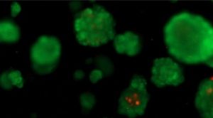
To develop effective probes for imaging complex tumor environments, single-chain polymer dots (Pdots) were synthesized using ultrasonic emulsification of D–A–D type conjugated polymers (Poly BT) with CTAB, resulting in highly cationic (+56.5 mV) and near-infrared (860 nm) emissive nanoparticles. These Pdots demonstrated excellent penetration into dense 3D tumor spheroids derived from MCF-7, SH-SY5Y, and PC-12 cell lines. Fluorescence imaging confirmed deep diffusion and mitochondrial colocalization, highlighting their strong potential as effective probes for bioimaging in complex tumor microenvironments.